


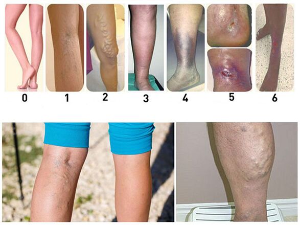
Varicose veins (varicose veins) is a disease in which superficial veins are dilated or swollen. The disease in most cases occurs in people older than 30 years. In the vast majority of cases, it is observed in the lower extremities. Varicose veins are characterized by dilation of the lumen of the veins with a simultaneous change in their wall. Saphenous veins are well formed, the direction of their flow becomes "snake-like". The large saphenous vein is usually affected, less often the small vein, and even less frequently their anastomoses.
Causes of varicose veins
The theories proposed to explain the causes and mechanisms of disease can be reduced to three groups.
The theories of the first group explain the formation of varicose veins by the anatomical characteristics of the location and structure of these vessels of the lower extremities. The veins have valves that prevent the centrifugal flow of blood, and thus its excessive flow from the subcutaneous to the deep veins of the legs. With insufficiency of the valves in the saphenous veins, more blood is deposited, which leads to their spread.
Theories of the second group in the development of varicose veins attach importance to the stagnation of blood in the pelvis during pregnancy, constipation, the consequences of inflammatory processes, as well as during a longer stay on your feet.
The theories of the third group are the least substantiated, which explain the origin of varicose veins by the constitutional predisposition, the weakness of the mesenchyme.
With varicose veins, for various reasons, their walls change, become thinner, and increased pressure leads to bulging of the walls. It first manifests itself in the form of nodules, and at the same time areas of compaction are observed as a result of excessive growth of connective tissue. Mechanical factors only contribute to the development of the pathological process in the veins, but they are in no way the main point of pathogenesis, etiology and causes of varicose veins of the lower extremities.
Symptoms of varicose veins
With varicose veins, patients usually have a feeling of fullness and heaviness in the lower extremities. Sometimes there is a short-lived, convulsive nature of the pain. There is often swelling. The feeling of fullness and heaviness in the limbs increases in the evening, because the edema usually increases by that time. Itching occurs, scratching of the legs often occurs. In the later stages of the disease, ulcers appear, which are usually located in the lower third of the lower leg on the inside.
The main objective symptom of the disease is visible varicose veins. Examination of the patient to determine this symptom is performed in a standing position. At the same time, varicose veins are clearly visible; on the lower leg they look more prominent, curved; on the thigh, the veins are usually dilated only along the main vascular trunk. Sometimes there is a varicose vein on the thigh almost at the mouth of the largest vein in the femoral vein. Such a nodule can be mistaken for a femoral hernia, but the softness of the nodule, its rapid filling with blood after taking the examiner's arm, and the presence of varicose veins on the lower leg are the basis for making a correct diagnosis.
There are a number of symptoms that indicate the presence of varicose veins of the great saphenous vein. This includes a symptom in which the patient is placed in a horizontal position and the leg is elevated. By carefully stroking the leg from the periphery to the center, the subcutaneous venous system is emptied, the place where the largest vein flows into the femoral vein is pressed firmly with the finger and holding the finger, the patient is transferred to a standing position. If the filling of the veins occurs only after the removal of the finger, then it is a positive symptom. In such cases, anastomoses between the superficial and deep venous networks are weakly expressed, and surgery can have a positive effect. If the patient's veins in the vertical position on the periphery start to fill slowly, it indicates a significant development of anastomosis - a negative symptom. In that case, the operation of ligation of the vein will not be successful.
The Delbe-Perthes symptom indicates how pronounced the discharge of the saphenous veins into the deep through the anastomoses is. An elastic bandage is placed on the patient in a standing position on the border of the middle and lower third of the thigh, and then they are offered to walk a little. If the tension of varicose veins decreases significantly, it indicates the presence of developed anastomoses between superficial and deep veins.
Other symptoms of varicose veins include swelling, eczematous skin changes and ulcers. Swelling is different - from mild pastiness to pronounced edema, when the skin loses its usual pattern and looks great, the circumference of the lower leg increases significantly. Eczematous manifestations include dryness, peeling and, finally, eczematous rash. The skin on the lower leg is usually affected. These changes occur as a result of trophic disorders.
Prevention and treatment of varicose veins
Prevention of varicose veins is reduced to a change of occupation, if it is associated with prolonged standing, taking measures for regular bowel movements, bandaging the legs with an elastic bandage or wearing elastic stockings. Bandaging the legs or wearing socks must be done lying down. For a few minutes, the leg is kept in an elevated position and only after making sure that the veins are empty, they put on a bandage or put on a sock. The bandage begins to be applied from below and continues upwards, avoiding any stretching and squeezing that causes stagnation.
There are a number of methods for surgical treatment. The operation of ligating the great saphenous vein into the Scarp triangle at the site of entry into the femoral vein is palliative. Relapses are often observed after this operation. Therefore, it is used only in combination with other surgical interventions.
During Bebcock's operation, a skin incision is made at the lower end of the varicose vein of the skin, it is separated and tied. A long abdominal probe is opened above the bend and inserted into the lumen. Another small incision in the skin is made above the upper end of the varicose vein. Its middle end is tied and crossed, below the section the vein is tightly tied over the probe, after which it is carefully removed through the lower incision. At the same time, the probe pulls the vein that the intima has turned upside down. The disadvantage of this method is that hematomas form at the site of broken anastomoses.
During Madelung surgery, varicose veins are excised throughout. Of all the surgeries, this intervention is the most radical and gives the best long-term results.
Complications of varicose veins
The most common and most difficult to treat complications of varicose veins are varicose ulcers. These ulcers usually occur in the elderly. They are located on the inner, less often on the outer surface of the lower third of the lower leg. These ulcers are the result of chronic tissue malnutrition. They are usually deep, have a necrotic bottom with an unpleasant-smelling discharge, and high, calloused edges. Ulcers can reach large sizes, rounding the entire lower leg. The skin around them is pigmented, sometimes inflamed, with eczematous irritation.
Varicose ulcers should be distinguished from syphilitic ones. Syphilitic ulcers are usually found in the upper third of the lower leg, more often on the front surface. In addition, other signs of syphilis may be found in syphilitic ulcers. Tuberculosis of the skin (lupus) is more common on the face, much less common on the extremities. Lupus begins as isolated nodules that then ulcerate; in the future there is deeper tissue damage, sometimes with the formation of smooth scars that tighten adjacent tissues.
Since varicose ulcers develop in the background of circulatory and trophic disorders, their treatment must be persistent and long-lasting. The constant position of the patient with a raised leg in most cases leads to a rapid improvement of the condition. The ulcer should be bandaged with a 0. 5% solution of potassium permanganate, with penicillin ointment or balsamic liniment. When the wound is cleansed and the swelling around it disappears, excision of the vein is recommended. Only radical surgery to remove the altered veins eliminates the risk of ulcer recurrence.
As the disease progresses and the enlarged nodules increase, their walls and the skin soldered to them become thinner. As a result, usually during walking (when the nodes are particularly tense), one of the nodes may rupture and venous bleeding may occur. Although such bleeding can be significant, it does not pose a great danger, because it stops quickly if the patient lies down and the leg is raised. In this position, negative pressure is created in the veins, they give way and the bleeding stops. A light aseptic bandage is applied to the wound. Due to the fact that bleeding can recur, surgery for excision of veins or their ligation and removal of the thinnest nodes is recommended. In case of bleeding from compensatory varicose veins, any operation related to ligation of the main vein trunk is categorically contraindicated.